Klicken Sie auf Einstellungen, falls Sie diese ablehnen möchten.
The Cardiological Heart Catheter Examination
The left and right heart catheters
Time to read: 4 Min.
Published in:
Diagnostics
at 26/07/2025
Cardiology Cardiac catheterisation is a diagnostic minimally invasive examination of the heart. This is performed by means of a coronary angiography when the presence of coronary heart disease is strongly suspected or to rule out coronary heart disease.
There are two types of cardiac catheterisation: The left-heart- catheter and the right heart catheter. The two examinations can be performed together or separately.
The Left Heart Catheter (also called arterial catheter or coronary angiography)
A thin tube is advanced to the heart via a hand or groin artery. Contrast medium is applied to the coronary vessels through this tube in order to be able to see an internal outflow of these vessels in the X-ray. This makes it possible to visualise constrictions. Coronary angiography is a diagnostic examination and is used to diagnose or exclude coronary heart disease (CHD). Coronary artery disease occurs when the coronary arteries show so-called calcifications, which narrow the vessels and prevent the heart muscle from being adequately supplied with blood and oxygen.
The typical clinical picture of this chronic condition is angina pectoris, which characteristically causes pain behind the breastbone and a feeling of tightness or constriction in the chest. However, the pain can also radiate to other areas. Acute blockage of a coronary artery is called infarction or heart attack.
Intervention
Coronary arteries that are narrowed or completely blocked can be dilated in an additional step with the help of a balloon placed on the catheter or drilled out (recanalised) with a wire. These areas are then kept open by inserting a stent. This minimally invasive procedure is called percutaneous transluminal coronary angioplasty" for short.
Angiography of other areas
In addition, other parts of the heart or vessels, such as the left ventricle, the heart valves (mitral or aortic valve) or the aorta, can be angiographed if necessary.
The Right Heart Catheter (also called venous catheter or right heart pressure measurement)
A right heart catheterisation is performed to assess the pressure in the pulmonary circulation. A thin tube is passed through the right heart via an arm, neck or groin vein into the vessels of the lung to measure the pressure there. This can be used to very accurately assess short-circuiting of the heart, the effects of valvular defects or heart failure.
Before the cardiac catheterisation
In the run-up to the examination, a detailed medical history and outpatient examination take place in the practice. If it turns out that a cardiac catheter examination is necessary, Dr Olaf Franzen explains the procedure to the patient in detail during a consultation.
The procedure of a cardiac catheterisation
The cardiac catheter examination is usually carried out by us in the cardiac catheter laboratory of the Hirslanden Klinik im Park in Zurich - as an outpatient or inpatient procedure, depending on the individual health situation.
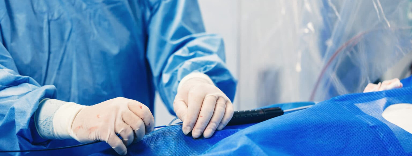
Preparation for the operation includes placing an intravenous line in your arm so that medication can be administered quickly in an emergency. In the cardiac catheterisation laboratory you will then be placed on the examination table under a large X-ray sheet and connected to a continuous ECG and blood pressure measurement. The groin or arm is then disinfected and locally anaesthetised for the vascular puncture. A general anaesthetic is not necessary.
The standard procedure today is to access the artery via the radial artery at the wrist. The femoral artery in the groin is now only punctured due to special conditions. After puncturing the vessel in question, a sheath is inserted through which the catheter is advanced to the heart. Radiological visualisation allows the doctor to assess the position of the catheter. The examination is not painful. Sometimes patients feel a slight pulling or a warm sensation when the contrast medium is injected.
The duration of the examination depends on the circumstances and the individual's health. A diagnostic cardiac catheterisation examination performed by a trained professional and free of complications usually takes a maximum of 15 to 30 minutes. If an additional intervention is necessary, the duration can sometimes be considerably longer.
After removal of the catheter and the sheath, the punctured area must be closed for several hours with a special pressure bandage to prevent secondary bleeding into the tissue. A puncture of the groin requires subsequent rest of the leg for several hours.
What must be observed after a cardiac catheter examination?
In order to avoid post-operative bleeding and complications, bed rest and rest is recommended - especially if the catheter was inserted through the groin. Excessive exertion and carrying heavy loads should be avoided for some time.
If an intervention has been performed, a stay in hospital for a few days may be necessary, depending on the extent of the intervention.
Anxiety (1)
Loss of appetite (1)
Arterial stenosis (3)
Breath sounds (1)
Shortness of Breath (5)
Balloon Dilatation (1)
Unconsciousness (2)
Anaemia (2)
Chest burn (1)
Chest constriction (2)
Chest pain (2)
Pressure measurement (1)
ECG (1)
Neck Vein Congestion (1)
Heart Attack (4)
Palpitations (1)
Cardiac Arrhythmia (2)
Pacemaker (0)
Heart Stumble (1)
Cardioversion (0)
Cardioverter (0)
Coronary Angiography (3)
Coronary Angioplasty (2)
Shortness of Breath (0)
LAA (1)
Weakness in Performance (3)
Mitraclip (2)
Fatigue (4)
Oedema (1)
PFO (0)
Coronary Angioplasty (1)
Irritant Cough (0)
Recanalisation (1)
REVEAL (0)
Red Head (0)
Stroke (3)
Pain Radiation (2)
Sweats (2)
Dizziness (2)
Stents (2)
TAVI (1)
TEE (1)
TTE (0)
Atrial Fibrillation (3)
Urination (2)